A study published in 2025 used a technique called autofluorescence imaging to look inside T cells before and after cryopreservation -- the standard freeze-and-store process used in most immune research labs and many commercial testing pipelines (https://pubmed.ncbi.nlm.nih.gov/42137595/). What the researchers found is relevant far beyond the laboratory bench. Frozen T cells did not behave like fresh ones. Their metabolic signals shifted, and their ability to activate -- to "turn on" in response to a threat -- was measurably blunted. That gap between the frozen version of your immune cells and the live version matters for how we interpret immune testing, and it matters for how we think about immune resilience in functional medicine.
This post explains what the research actually shows, why T cell metabolism is central to immune function, and what it means practically if you are trying to understand your own immune health.
What autofluorescence imaging actually does
Most people picture immune research as a process involving stains, dyes, or radioactive tracers -- something applied to cells from the outside so a camera can see them. Autofluorescence imaging works differently. It detects light that cells emit naturally, without any added marker.
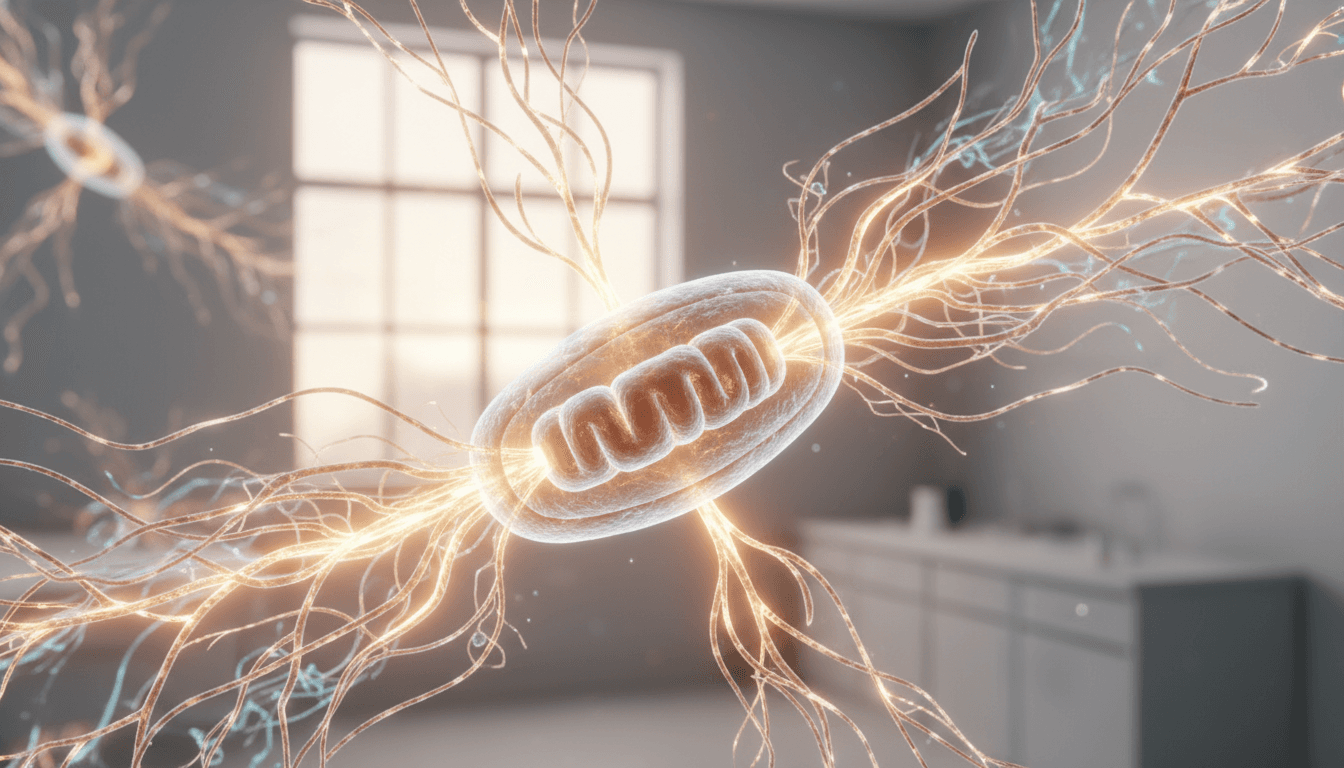
The signals come primarily from two coenzymes: NADH (the reduced form of NAD+) and FAD (flavin adenine dinucleotide). Both of these molecules are embedded in the mitochondrial energy-production process. When a cell is actively burning fuel through oxidative phosphorylation -- the high-efficiency energy pathway that powers most resting and memory immune functions -- the ratio of NADH to FAD shifts in a measurable direction (https://doi.org/10.1038/srep01936). Researchers call this ratio the optical redox ratio, and it functions as a real-time readout of metabolic activity without disturbing the cell at all.
This is not a new concept in cancer biology -- autofluorescence has been used to detect metabolic signatures in breast tissue for over a decade (https://doi.org/10.1038/srep01936). What is newer is applying it systematically to immune cells, specifically to ask: does the way we handle T cells before we test them change what we see?
The 2025 study answered that question with a clear yes. Cryopreserved T cells showed altered optical redox ratios compared with freshly processed cells, and they mounted a weaker metabolic response when stimulated with an activation signal. In plain terms: the frozen cells had a different energy fingerprint, and they were slower to mobilize (https://pubmed.ncbi.nlm.nih.gov/42137595/).
Why T cell metabolism is not a niche topic
It is tempting to file this under "interesting science, not relevant to me." But T cell metabolism sits at the center of immune function in ways that touch everyday clinical questions.
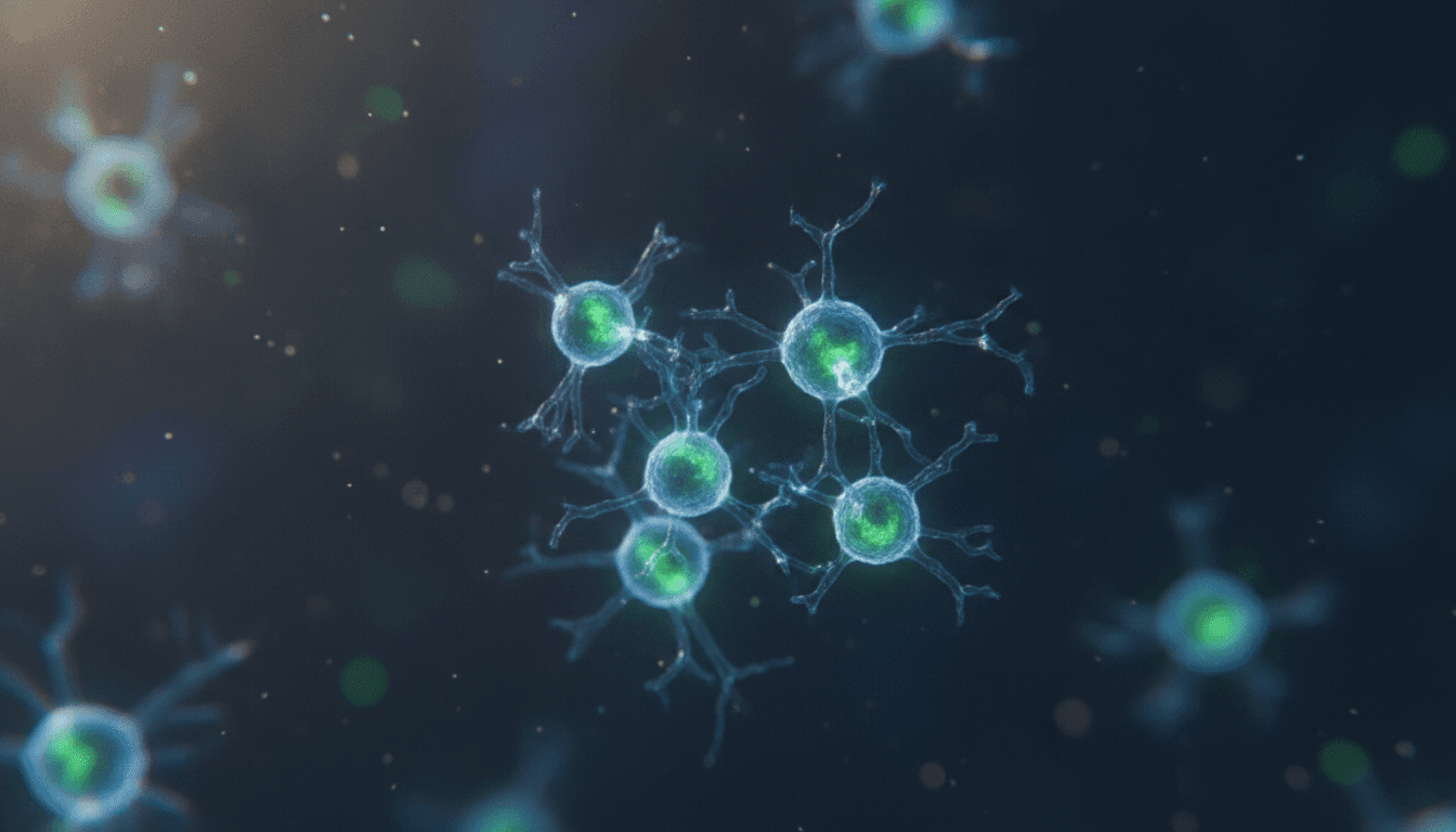
T cells are the coordinators of adaptive immunity. They identify infected or abnormal cells, direct the inflammatory response, and build the memory that lets your immune system respond faster to a second exposure. What determines how well they do all of that? Energy (https://doi.org/10.1126/science.1242454).
A naive T cell -- one that has not encountered a threat yet -- runs primarily on oxidative phosphorylation, the slow-burn, high-efficiency mitochondrial pathway. When it receives an activation signal, it has to shift rapidly into glycolysis, a faster but less efficient pathway that generates the ATP needed for rapid cell division and cytokine production. This switch is called metabolic reprogramming, and it is not optional. A T cell that cannot make the switch cannot mount a proper immune response (https://doi.org/10.1016/j.cell.2017.04.004).
After the immediate threat passes, the T cells that survive and become memory cells shift back toward oxidative phosphorylation -- specifically a high-capacity mitochondrial state that allows them to persist for years and respond quickly to a future encounter (https://doi.org/10.1016/j.immuni.2011.12.007). The mitochondrial health of those memory T cells is a major determinant of long-term immune resilience.
This is where the research connects to functional medicine. The same inputs that support mitochondrial function across the body -- adequate NAD+ availability, quality sleep, controlled inflammation, and exercise -- also support the metabolic readiness of your T cells. And the same stressors that impair cellular energy production -- chronic sleep deprivation, persistent low-grade inflammation, sedentary behavior, excess alcohol -- impair T cell metabolism too.
What cryopreservation does to the cell's energy state
Cryopreservation is not inherently damaging in the gross sense. Properly frozen and thawed T cells are still alive, still structurally recognizable, and still capable of performing basic immune functions. That is why it has become the standard in large-scale clinical trials and commercial immune profiling -- samples from different sites can be batch-processed in a central laboratory, which controls for inter-assay variability.
The problem the 2025 study identified is subtler. The freeze-thaw cycle imposes a stress on mitochondria. Ice crystal formation, osmotic shifts during the thaw, and cold-induced metabolic depression all leave a fingerprint on the cell's energy state even after the cell has technically "recovered" (https://pubmed.ncbi.nlm.nih.gov/42137595/). Autofluorescence imaging is sensitive enough to detect that fingerprint where simpler viability assays -- which only ask "alive or dead?" -- would miss it entirely.
The practical implication is that a commercial immune panel run on a cryopreserved sample may be measuring a slightly different cell than the one that is actually circulating in your bloodstream today. For most routine markers, the difference is small and the validated reference ranges already account for it. For more nuanced metabolic or functional immune assessments, the gap matters more. We think that is worth knowing -- and worth asking about when you review your results with a clinician.
What this means for how we interpret immune testing
We are not saying immune testing is unreliable. We are saying interpretation requires context, which is true of every test in medicine.
What clinicians look for when evaluating immune function goes well beyond a single frozen snapshot. A thorough functional immune workup considers the full clinical picture: chronic infection history, inflammatory markers, sleep data, stress load, nutritional status, and the trajectory of results over time. A single data point from a cryopreserved sample is useful input. It is not a verdict.
The autofluorescence research adds a useful layer of nuance. It is a reminder that the way a sample is handled between the draw and the analysis introduces variables -- and that as imaging and metabolic profiling tools become more accessible, we will get better at detecting those variables rather than averaging over them.
For anyone interested in the broader landscape of cellular energy and immune health, our post on functional wellness without the woo covers how we think about evidence quality in this space. And if you are curious specifically about NAD+ and mitochondrial support -- which overlaps directly with the T cell metabolism research here -- our NAD therapy post walks through what the human evidence actually shows.
The NAD+ connection -- and what the evidence supports
NADH is not just a fluorescence signal. It is one of the most important electron carriers in the cell. Its availability determines how efficiently mitochondria can run oxidative phosphorylation, and by extension, how well T cells can sustain the high-energy state needed for immune memory.
NAD+ -- the oxidized form -- is the precursor. Adequate NAD+ availability allows cells to regenerate NADH on demand during active metabolism. In aging, chronic inflammation, and metabolic stress, NAD+ levels decline, and cellular energy capacity declines with them (https://doi.org/10.1101/sqb.2011.76.010439).
Preclinical data and early human studies have explored whether supplementing NAD+ precursors -- nicotinamide riboside (NR) and nicotinamide mononucleotide (NMN) being the most studied -- can restore cellular NAD+ levels and functional outcomes. The evidence is promising in specific contexts but still developing in humans. We are honest about that distinction: interesting mechanism does not automatically equal proven clinical outcome, and we discuss that boundary carefully with every patient before recommending any supplement protocol.
What is clear is that the mitochondrial framework the autofluorescence research uses to evaluate T cells is the same framework that connects sleep quality, exercise capacity, metabolic health, and immune resilience across the body. These are not separate conversations. They are the same conversation at different scales.
Practical takeaways from a research-forward functional medicine lens
So what does this all add up to for someone sitting in a clinic -- or reading this at home -- trying to understand their immune health?
First, ask questions about your immune testing methodology. If your results come from a frozen sample processed at a central lab, that is normal and usually fine -- but knowing it helps you interpret outliers more accurately. If a result looks unexpectedly low, methodology is one variable worth checking before assuming a pathological cause.
Second, pay attention to the inputs that support T cell metabolic readiness. The evidence-based list is not glamorous, but it is consistent across the immunology literature: consistent, restorative sleep (the mitochondrial cleanup window), adequate dietary protein to support lymphocyte turnover, resistance training to maintain oxidative capacity in immune and muscle tissue, and management of chronic low-grade inflammation through diet quality and stress modulation (https://doi.org/10.1146/annurev-immunol-042617-053019).
Third, if you are interested in deeper immune or metabolic profiling -- the kind that goes beyond a standard CBC differential -- that conversation is worth having with a clinician who understands the methodological nuances. The tools are improving quickly. What autofluorescence imaging can do in a research setting today will likely be part of more accessible clinical workflows within the decade.
For context on how chronic systemic inflammation connects to these immune questions, our post on multisystemic inflammation covers the clinical picture from a different angle.
Why this research matters beyond the lab bench
Science moves from bench to bedside slowly, and a single imaging study does not change clinical practice overnight. But the autofluorescence research matters for a few reasons that go beyond its immediate findings.
It demonstrates that standard sample-handling protocols -- ones we have used for decades -- can introduce biologically meaningful variation in how immune cells look and behave. That is a methodological signal for the entire field of immune profiling. It also validates the optical redox ratio as a practical, label-free readout of immune cell metabolic state, which opens doors for non-invasive monitoring approaches that do not require freezing samples at all.
And it reinforces something we think about constantly in functional medicine: that the gap between "technically normal" and "optimally functional" is real, measurable, and worth caring about. A T cell that is alive is not the same as a T cell that is metabolically ready. The same logic applies to every tissue in the body.
If you want to explore your immune health, metabolic function, or the full functional medicine picture in a setting where results are interpreted in clinical context -- not just compared to a population average -- we would like to talk. Book a visit with us at our functional medicine page, or call NoMi Beach Health at (786) 744-5152. Dr. Jezwah Harris (NP, JD, MBA, FNP-BC, MEP-C) will review your history, your labs, and your goals, and give you a straight answer about what the data actually means for you.
Frequently Asked Questions
- What is autofluorescence imaging and why does it matter for immune testing?
- Autofluorescence imaging measures naturally occurring light signals from metabolic molecules inside cells -- no dyes needed. Researchers use it to assess how active a cell's energy machinery is. For immune testing, this matters because a cell that looks structurally intact under a standard microscope can still be metabolically impaired, and autofluorescence catches that difference.
- What is cryopreservation and why is it used with T cells?
- Cryopreservation means freezing biological samples -- including blood cells -- at very low temperatures so they can be stored and shipped to a laboratory without immediate processing. It is standard practice in clinical trials and many commercial immune panels. The trade-off is that the freeze-thaw process can stress or alter the cells being studied.
- Does this research mean my immune lab results from a frozen sample are wrong?
- Not necessarily wrong, but possibly shifted from what a fresh sample would show. The study found that cryopreserved T cells showed altered metabolic signatures and blunted activation responses compared with fresh cells. What that means clinically depends heavily on the specific test and how your lab accounts for this variation.
- What are T cells and why do clinicians care about their metabolic state?
- T cells are a class of white blood cells that coordinate immune defense -- attacking infected cells, regulating inflammation, and building immunological memory. Their metabolic state tells clinicians how ready those cells are to respond. A T cell that cannot shift into high-energy mode when needed is less effective, which is relevant in chronic infections, autoimmune conditions, and recovery from illness.
- How does T cell metabolism connect to the broader functional wellness picture?
- T cell metabolism depends on the same cellular energy systems -- primarily mitochondrial oxidative phosphorylation and glycolysis -- that underlie fatigue, inflammation, and resilience across the whole body. Factors like NAD+ availability, sleep quality, and chronic stress all influence how well immune cells can fuel themselves and respond to threats.
- Should I ask for fresh-sample immune testing instead of frozen?
- That is a conversation worth having with your clinician. Fresh-sample processing removes the cryopreservation variable but requires on-site or same-day laboratory capacity. For many routine panels, standard frozen protocols are well-validated. For research-grade metabolic immune profiling, the choice of protocol matters more and should be discussed with whoever is interpreting your results.
- What practical steps support T cell metabolic health right now?
- The evidence base points to consistent, quality sleep, management of chronic low-grade inflammation, adequate protein intake, resistance exercise, and -- where clinically indicated -- addressing nutrient gaps such as NAD+ precursors and vitamin D. None of these are novel, but the immune-metabolic research gives them a more precise mechanistic rationale.
Sources
- Shah S, et al. Autofluorescence imaging reveals the impact of cryopreservation on T cell metabolism and activation response. bioRxiv (2025).
- Buck MD, Sowell RT, Kaech SM, Pearce EL. Metabolic instruction of immunity. Cell (2017).
- Pearce EL, Poffenberger MC, Chang CH, Jones RG. Fueling immunity: insights into metabolism and lymphocyte function. Science (2013).
- Quinn KP, et al. Quantitative metabolic imaging using endogenous fluorescence to detect signatures of breast cancer. Sci Rep (2013).
- Galganov Y, et al. Optical metabolic imaging of T cells identifies metabolic reprogramming during activation. PLOS ONE (2022).
- Nicholson LB. The immune system. Essays Biochem (2016).
- Crompton JG, et al. Akt inhibition enhances expansion of potent tumor-specific lymphocytes with memory cell characteristics. Cancer Res (2015).
- van der Windt GJ, et al. Mitochondrial respiratory capacity is a critical regulator of CD8+ T cell memory development. Immunity (2012).
- Cantó C, Auwerx J. NAD+ as a signaling molecule modulating metabolism. Cold Spring Harb Symp Quant Biol (2011).
- Prlic M, Bevan MJ. Immunology: a metabolic switch to memory. Nature (2009).
- Warburg O, Wind F, Negelein E. The metabolism of tumors in the body. J Gen Physiol (1927).
- Geltink RIK, Kyle RL, Pearce EL. Unraveling the complex interplay between T cell metabolism and function. Annu Rev Immunol (2018).